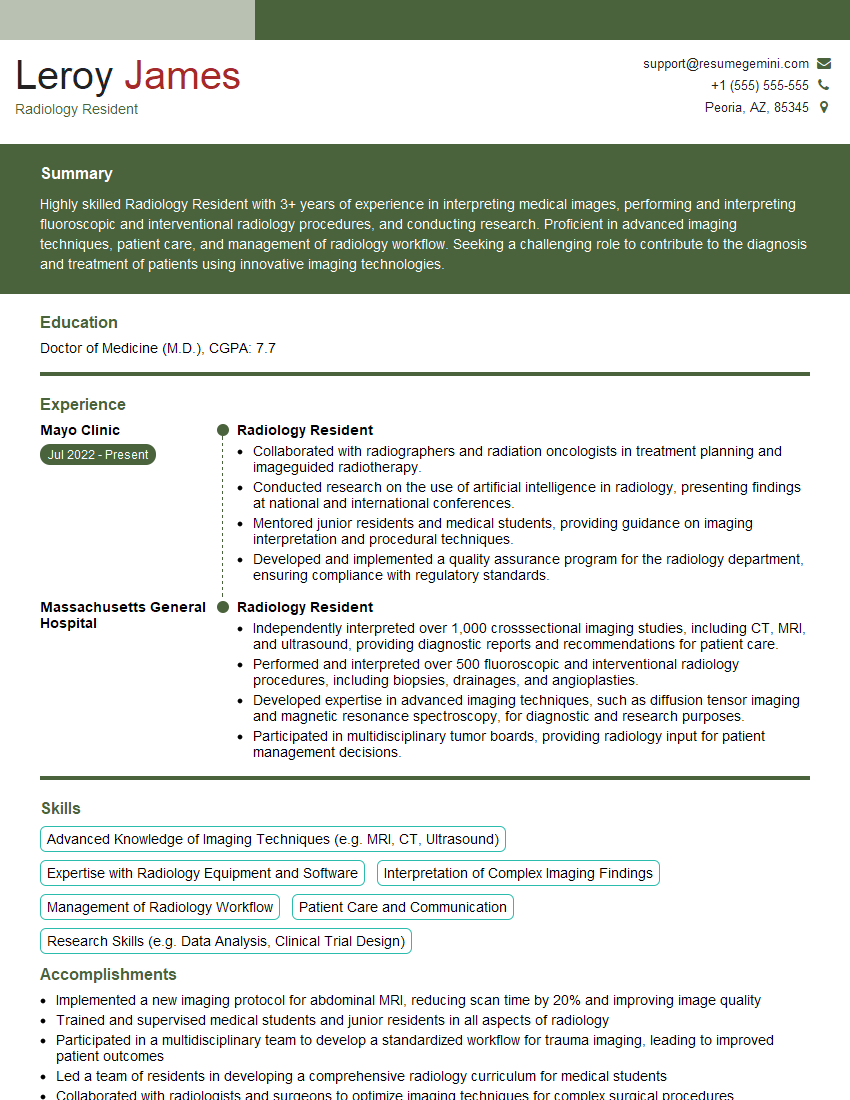
Feeling lost in a sea of interview questions? Landed that dream interview for Radiology Resident but worried you might not have the answers? You’re not alone! This blog is your guide for interview success. We’ll break down the most common Radiology Resident interview questions, providing insightful answers and tips to leave a lasting impression. Plus, we’ll delve into the key responsibilities of this exciting role, so you can walk into your interview feeling confident and prepared.
Acing the interview is crucial, but landing one requires a compelling resume that gets you noticed. Crafting a professional document that highlights your skills and experience is the first step toward interview success. ResumeGemini can help you build a standout resume that gets you called in for that dream job.
Essential Interview Questions For Radiology Resident
1. Describe the key factors you consider when evaluating a chest X-ray for a patient with shortness of breath?
When evaluating a chest X-ray for a patient with shortness of breath, I consider the following key factors:
- Cardiac size and shape: Cardiomegaly or an abnormal cardiac silhouette may indicate heart failure or pericardial effusion.
- Pulmonary vascularity: Increased or decreased pulmonary vascularity can suggest pulmonary edema, interstitial lung disease, or pulmonary embolism, respectively.
- Lung parenchyma: Infiltrates, consolidation, or nodules may indicate pneumonia, pulmonary edema, or lung cancer.
- Pleural space: Pleural effusions, pneumothorax, or pleural thickening may cause shortness of breath.
- Diaphragm and mediastinum: A high or low diaphragm, mediastinal widening, or masses can indicate diaphragmatic dysfunction, aortic dissection, or other mediastinal pathology.
2. How do you approach interpreting a CT scan of the abdomen and pelvis in a patient with suspected appendicitis?
Window settings
- Use abdominal and pelvic windows to optimize visualization of different structures.
Appendiceal anatomy
- Identify the location of the appendix, usually in the right lower quadrant.
- Note its size, shape, and relationship to surrounding structures.
Signs of appendicitis
- Look for thickening and enhancement of the appendix wall.
- Check for obliteration of the appendiceal lumen.
- Evaluate for surrounding inflammation, such as peri-appendiceal fat stranding or abscess formation.
Differential diagnoses
- Consider other conditions that can mimic appendicitis, such as Meckel’s diverticulitis or right-sided colonic diverticulitis.
3. What are the imaging findings suggestive of a brain tumor in a patient presenting with seizures?
- Mass lesion: A focal area of abnormal tissue density, typically iso- or hyperintense on T2 and heterogeneously enhancing on gadolinium.
- Edema: Surrounding edema, causing mass effect and distortion of normal brain structures.
- Abnormal enhancement: Irregular or nodular enhancement, often with a central necrotic core.
- Contrast enhancement: Peripheral or ring-like enhancement on post-contrast images.
- Diffusion restriction: Reduced diffusion of water molecules within the tumor, indicating cellularity or high grade.
- Spectroscopy: Metabolic changes, such as elevated choline or reduced NAA, can suggest malignancy.
4. How do you evaluate a patient with suspected pancreatitis on an abdominal MRI?
- T1-weighted images: Look for pancreatic enlargement, edema, and fluid collections.
- T2-weighted images: Assess for increased pancreatic and peripancreatic signal, which may indicate inflammation and edema.
- Fat-suppressed T2-weighted images: Improve visualization of the pancreas and surrounding structures.
- Contrast-enhanced images: Evaluate for pancreatic ductal dilatation, pseudocyst formation, and areas of delayed enhancement, which can suggest necrosis.
- MR cholangiopancreatography (MRCP): Visualize the pancreatic and biliary ducts to assess for stones, strictures, or other abnormalities.
5. Describe the imaging features of a vertebral compression fracture on plain radiographs?
- Loss of vertebral height: Reduction in the vertical dimension of the affected vertebra.
- Increased vertebral density: Condensation of the trabecular bone, resulting in a brighter appearance.
- Angular deformity: A wedge-shaped appearance of the vertebral body, with the anterior height being less than the posterior height.
- Posterior vertebral body margin irregularity: A smooth or step-like deformity of the posterior vertebral body margin, known as the “posterior vertebral body depression” sign.
- Paravertebral soft tissue swelling: Associated edema or hematoma may be visible in acute fractures.
6. How do you use ultrasound to evaluate the thyroid gland?
- Transverse and longitudinal views: Visualize the size, shape, and echo-texture of the thyroid lobes.
- Nodule evaluation: Assess the size, shape, margins, and echogenicity of thyroid nodules.
- Blood flow: Use color Doppler ultrasound to evaluate intrathyroidal blood flow, which can help differentiate benign from malignant nodules.
- Lymph node assessment: Examine the cervical lymph nodes for enlargement or abnormal echogenicity.
7. Describe the role of nuclear medicine in evaluating a patient with suspected myocardial infarction?
- Myocardial perfusion imaging: Assess for regional myocardial ischemia or infarction by injecting a radioactive tracer and imaging the heart at rest and during stress.
- SPECT (single-photon emission computed tomography): Provides three-dimensional images of myocardial perfusion, allowing for better localization and quantification of ischemic areas.
- PET (positron emission tomography): Uses a radioactive tracer that emits positrons, allowing for more specific imaging of myocardial viability and metabolism.
8. How do you interpret a bone scan in a patient with suspected metastatic disease?
- Increased uptake: Focal areas of increased tracer uptake can indicate metastatic lesions or other bone abnormalities.
- Multiple lesions: Widespread involvement of multiple bones suggests disseminated metastatic disease.
- Pattern of uptake: Different patterns, such as lytic, sclerotic, or mixed, can provide clues to the underlying etiology.
- Correlation with other imaging: Combine bone scan findings with plain radiographs, CT, or MRI for a comprehensive assessment.
9. What are the key imaging features that differentiate acute from chronic renal failure on ultrasound?
Acute renal failure
- Increased renal parenchymal echogenicity: Swollen and edematous kidneys appear brighter than normal.
- Loss of corticomedullary differentiation: The normal distinction between the cortex and medulla becomes less clear.
- Increased renal size: The kidneys may be enlarged due to edema.
Chronic renal failure
- Small, contracted kidneys: Shrunken kidneys with decreased size and increased echogenicity.
- Loss of renal contour: Smooth and rounded kidney outlines.
- Increased parenchymal heterogeneity: Patchy areas of increased and decreased echogenicity throughout the kidneys.
10. Discuss the role of contrast-enhanced CT in evaluating a patient with suspected pulmonary embolism?
- Pulmonary artery visualization: Intravenous contrast injection allows for clear visualization of the pulmonary arteries, enabling detection of emboli.
- Filling defects: Pulmonary emboli appear as filling defects within the enlarged pulmonary arteries.
- Central vs. peripheral embolism: CT can differentiate between central (main pulmonary artery or lobar branches) and peripheral (segmental or subsegmental branches) emboli.
- Collateral vessels: Contrast-enhanced CT can reveal collateral vessels that develop in response to chronic emboli.
- Alternative diagnoses: CT also allows for evaluation of other potential causes of dyspnea, such as pneumonia, lung masses, or pleural effusions.
Interviewers often ask about specific skills and experiences. With ResumeGemini‘s customizable templates, you can tailor your resume to showcase the skills most relevant to the position, making a powerful first impression. Also check out Resume Template specially tailored for Radiology Resident.
Career Expert Tips:
- Ace those interviews! Prepare effectively by reviewing the Top 50 Most Common Interview Questions on ResumeGemini.
- Navigate your job search with confidence! Explore a wide range of Career Tips on ResumeGemini. Learn about common challenges and recommendations to overcome them.
- Craft the perfect resume! Master the Art of Resume Writing with ResumeGemini’s guide. Showcase your unique qualifications and achievements effectively.
- Great Savings With New Year Deals and Discounts! In 2025, boost your job search and build your dream resume with ResumeGemini’s ATS optimized templates.
Researching the company and tailoring your answers is essential. Once you have a clear understanding of the Radiology Resident‘s requirements, you can use ResumeGemini to adjust your resume to perfectly match the job description.
Key Job Responsibilities
Radiology Residents are responsible for providing comprehensive radiological care to patients under the supervision of attending radiologists. They perform a wide range of diagnostic imaging procedures, including X-rays, CT scans, MRIs, and ultrasound exams. Residents also interpret medical images and provide written reports to referring physicians. Additionally, they may participate in research projects and teaching activities.
1. Diagnostic Imaging
Residents perform a variety of diagnostic imaging procedures, including:
- X-rays
- CT scans
- MRIs
- Ultrasound exams
2. Image Interpretation
Residents interpret medical images and provide written reports to referring physicians. These reports describe the findings of the imaging examination and may include recommendations for further evaluation or treatment.
- Reviewing and interpreting images from various imaging modalities
- Identifying and describing abnormalities and findings
- Correlating findings with clinical history and physical examination
3. Patient Care
Residents provide patient care under the supervision of attending radiologists. This includes:
- Explaining procedures to patients
- Positioning patients for exams
- Monitoring patients during exams
4. Education and Research
Residents may participate in research projects and teaching activities. This may include:
- Conducting clinical research studies
- Presenting research findings at conferences
- Teaching medical students and other healthcare professionals
Interview Tips
Preparing for a Radiology Residency interview can be a daunting task, but following certain tips and strategies can help you put your best foot forward and increase your chances of success.
1. Research the Program
Before the interview, take some time to research the Radiology Residency program you are applying to. This will help you understand the program’s history, mission, and goals. You should also familiarize yourself with the faculty and their research interests. This will help you demonstrate your interest in the program and ask informed questions during the interview.
- Visit the program’s website
- Read the program’s mission statement
- Review the faculty’s profiles
- Attend a program information session
2. Practice Your Answers
It is likely that you will be asked a number of common interview questions. Take some time to practice your answers to these questions. This will help you feel more confident and prepared during the interview. Common interview questions include:
- “Tell me about yourself.”
- “Why are you interested in this program?”
- “What are your strengths and weaknesses?”
- “What are your research interests?”
- “What are your career goals?”
3. Dress Professionally
First impressions matter, so it is important to dress professionally for your interview. This means wearing a suit or business casual attire. You should also make sure that your clothes are clean and pressed. Avoid wearing clothes that are too revealing or too casual.
4. Be Yourself
It is important to be yourself during your interview. The interviewers want to get to know the real you. So be honest and genuine in your answers. Don’t try to be someone you’re not. The interviewers will be able to tell if you’re being fake.
5. Follow Up
After the interview, be sure to follow up with a thank-you note. This is a simple way to show your appreciation for the interviewers’ time and to reiterate your interest in the program. In your note, you can also mention any specific aspects of the program or the interview that you found particularly interesting.
Next Step:
Now that you’re armed with a solid understanding of what it takes to succeed as a Radiology Resident, it’s time to turn that knowledge into action. Take a moment to revisit your resume, ensuring it highlights your relevant skills and experiences. Tailor it to reflect the insights you’ve gained from this blog and make it shine with your unique qualifications. Don’t wait for opportunities to come to you—start applying for Radiology Resident positions today and take the first step towards your next career milestone. Your dream job is within reach, and with a polished resume and targeted applications, you’ll be well on your way to achieving your career goals! Build your resume now with ResumeGemini.
