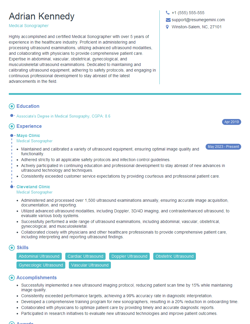
Are you gearing up for a career in Medical Sonographer? Feeling nervous about the interview questions that might come your way? Don’t worry, you’re in the right place. In this blog post, we’ll dive deep into the most common interview questions for Medical Sonographer and provide you with expert-backed answers. We’ll also explore the key responsibilities of this role so you can tailor your responses to showcase your perfect fit.
Acing the interview is crucial, but landing one requires a compelling resume that gets you noticed. Crafting a professional document that highlights your skills and experience is the first step toward interview success. ResumeGemini can help you build a standout resume that gets you called in for that dream job.
Essential Interview Questions For Medical Sonographer
1. Describe the key components of the ultrasound system, and explain their functions.
The key components of an ultrasound system are:
- Transducer: The transducer is the probe that is placed on the patient’s body. It emits ultrasound waves and receives the reflected echoes.
- Signal processing unit: The signal processing unit processes the echoes received from the transducer and creates an image of the scanned area.
- Display unit: The display unit displays the ultrasound image on a monitor.
- Control panel: The control panel allows the user to control the ultrasound system settings, such as the depth of the scan and the frequency of the ultrasound waves.
2. What are the different types of ultrasound transducers, and what are their applications?
Linear transducer
- Used for superficial imaging, such as imaging the thyroid, breast, and muscles.
- Provides high-resolution images with good tissue penetration.
Curved transducer
- Used for imaging deeper structures, such as the abdomen and heart.
- Provides a wider field of view than a linear transducer.
Phased array transducer
- Used for 3D imaging and Doppler imaging.
- Allows for real-time imaging of moving structures, such as the heart and blood vessels.
3. Describe the different modes of ultrasound imaging, and explain their advantages and disadvantages.
The main modes of ultrasound imaging are:
- B-mode (brightness mode): Produces a 2D image of the scanned area. The brightness of each pixel in the image corresponds to the strength of the reflected echoes.
- M-mode (motion mode): Produces a 1D image of the movement of a structure over time. This mode is used to assess cardiac function.
- Doppler mode: Measures the velocity of blood flow in vessels. This mode is used to diagnose and assess the severity of vascular diseases.
- 3D mode: Produces a 3D image of the scanned area. This mode is used for complex anatomical structures, such as the heart and fetal structures.
4. What are the most common artefacts encountered in ultrasound imaging, and how can they be minimised?
The most common artefacts encountered in ultrasound imaging are:
- Reverberation artefact: Caused by multiple reflections of the ultrasound waves within the body. Can be minimised by using a lower frequency transducer.
- Shadowing artefact: Caused by the attenuation of the ultrasound waves by a dense structure, such as bone or air. Can be minimised by using a higher frequency transducer.
- Sidelobe artefact: Caused by the secondary lobes of the ultrasound beam. Can be minimised by using a transducer with a narrower beam width.
- Motion artefact: Caused by the movement of the patient or the transducer during the scan. Can be minimised by using a higher frame rate.
5. What are the safety considerations for ultrasound imaging, and how can they be ensured?
The safety considerations for ultrasound imaging include:
- Thermal effects: Ultrasound waves can cause a rise in temperature in the body. This can be minimised by using a lower power setting and avoiding prolonged exposure.
- Mechanical effects: Ultrasound waves can cause cavitation (the formation of bubbles in the body). This can be minimised by using a lower frequency transducer and avoiding prolonged exposure.
- Biological effects: Ultrasound waves can have a biological effect on cells. However, these effects are generally not harmful at the levels used in diagnostic ultrasound imaging.
- Patient safety: It is important to ensure that the patient is comfortable and well-informed about the procedure. The patient should be given the opportunity to ask questions and express any concerns.
6. What is your experience with using contrast agents in ultrasound imaging?
Contrast agents are used in ultrasound imaging to enhance the visibility of certain structures or to assess blood flow. I have experience using both microbubble contrast agents and sulphur hexafluoride microbubbles.
- Microbubble contrast agents: These are small, gas-filled bubbles that are injected into the bloodstream. They can be used to enhance the visibility of blood vessels and to assess blood flow.
- Sulphur hexafluoride microbubbles: These are small, gas-filled bubbles that are used to enhance the visibility of the left ventricular myocardium.
7. What is the role of the medical sonographer in the diagnosis and management of disease?
Medical sonographers play a vital role in the diagnosis and management of disease. They provide images that can be used to diagnose a wide range of conditions, including:
- Heart disease
- Cancer
- Abdominal and pelvic disorders
- Musculoskeletal disorders
- Vascular disorders
Medical sonographers also play a role in guiding interventional procedures, such as biopsies and needle aspirations.
8. What are the ethical considerations for medical sonographers?
Medical sonographers have a number of ethical considerations to be aware of, including:
- Patient confidentiality: Medical sonographers must maintain the confidentiality of patient information.
- Informed consent: Medical sonographers must ensure that patients understand the procedure and the risks involved before giving their consent.
- Professionalism: Medical sonographers must maintain a high level of professionalism at all times.
- Competence: Medical sonographers must only perform procedures that they are competent to perform.
9. What are your strengths and weaknesses as a medical sonographer?
Strengths:
- Excellent technical skills: I have a strong understanding of the principles of ultrasound imaging and am proficient in using all types of ultrasound equipment.
- Good communication skills: I am able to communicate effectively with patients and other members of the healthcare team.
- Patient-centred care: I am passionate about providing high-quality care to patients and am always looking for ways to improve my skills and knowledge.
Weaknesses:
- Limited experience with contrast agents: I have only used contrast agents a few times, so I would like to gain more experience in this area.
- Can be a bit of a perfectionist: I sometimes spend too much time trying to get the perfect image, which can slow down the workflow.
10. What are your career goals as a medical sonographer?
My career goal is to become a highly skilled and experienced medical sonographer. I am particularly interested in using ultrasound to diagnose and manage cardiovascular disease. I believe that my technical skills and patient-centred approach will make me a valuable asset to any healthcare team.
Interviewers often ask about specific skills and experiences. With ResumeGemini‘s customizable templates, you can tailor your resume to showcase the skills most relevant to the position, making a powerful first impression. Also check out Resume Template specially tailored for Medical Sonographer.
Career Expert Tips:
- Ace those interviews! Prepare effectively by reviewing the Top 50 Most Common Interview Questions on ResumeGemini.
- Navigate your job search with confidence! Explore a wide range of Career Tips on ResumeGemini. Learn about common challenges and recommendations to overcome them.
- Craft the perfect resume! Master the Art of Resume Writing with ResumeGemini’s guide. Showcase your unique qualifications and achievements effectively.
- Great Savings With New Year Deals and Discounts! In 2025, boost your job search and build your dream resume with ResumeGemini’s ATS optimized templates.
Researching the company and tailoring your answers is essential. Once you have a clear understanding of the Medical Sonographer‘s requirements, you can use ResumeGemini to adjust your resume to perfectly match the job description.
Key Job Responsibilities
Medical Sonographers are highly skilled healthcare professionals who use ultrasound technology to create images of the body’s internal organs and structures. They play a vital role in the diagnosis and treatment of various medical conditions.
1. Patient care
Providing excellent patient care is paramount. This includes greeting patients, explaining procedures, and ensuring their comfort throughout the examination.
- Maintaining a clean and safe work environment.
- Providing clear and concise instructions to patients.
2. Image acquisition
Medical Sonographers are responsible for obtaining high-quality images. This involves operating ultrasound equipment and adjusting settings to optimize image quality.
- Performing various ultrasound exams, such as abdominal, cardiac, and obstetric.
- Using different ultrasound probes and techniques to capture images from various angles.
3. Image interpretation
Medical Sonographers are trained to interpret ultrasound images and identify abnormalities. They collaborate with physicians to provide diagnostic information.
- Identifying anatomical structures and assessing their appearance.
- Recognizing signs of disease or injury.
4. Report generation
Medical Sonographers prepare written reports that summarize the findings of ultrasound examinations. These reports are used by physicians to make informed decisions about patient care.
- Documenting patient information, exam details, and observations.
- Communicating findings clearly and accurately.
Interview Tips
Preparing thoroughly for an interview can significantly increase your chances of success. Here are some essential tips to help you ace your Medical Sonographer interview:
1. Research the company and position
Demonstrating knowledge of the healthcare organization and the specific role you’re applying for shows your interest and enthusiasm. Research the company’s mission, values, and recent developments.
- Visit the company’s website and social media pages.
- Review the job description carefully and identify the key requirements.
2. Practice your answers to common interview questions
Anticipating and preparing for common interview questions can boost your confidence and help you deliver clear and concise responses.
- Prepare a brief introduction that highlights your qualifications and experience.
- Practice answering questions about your technical skills, patient care experience, and teamwork abilities.
3. Showcase your passion for medical imaging
Conveying your genuine interest in medical imaging and patient care is crucial. Share examples that demonstrate your commitment to the field.
- Describe a time when you went above and beyond to help a patient or provide exceptional customer service.
- Explain why you chose to pursue a career in medical sonography and what motivates you.
4. Dress professionally and arrive on time
First impressions matter. Dressing appropriately and arriving punctually for your interview demonstrate respect for the interviewer and the organization.
- Choose business attire that is clean, pressed, and fits well.
- Plan your route and allow ample time for travel and parking.
Next Step:
Armed with this knowledge, you’re now well-equipped to tackle the Medical Sonographer interview with confidence. Remember, preparation is key. So, start crafting your resume, highlighting your relevant skills and experiences. Don’t be afraid to tailor your application to each specific job posting. With the right approach and a bit of practice, you’ll be well on your way to landing your dream job. Build your resume now from scratch or optimize your existing resume with ResumeGemini. Wish you luck in your career journey!
