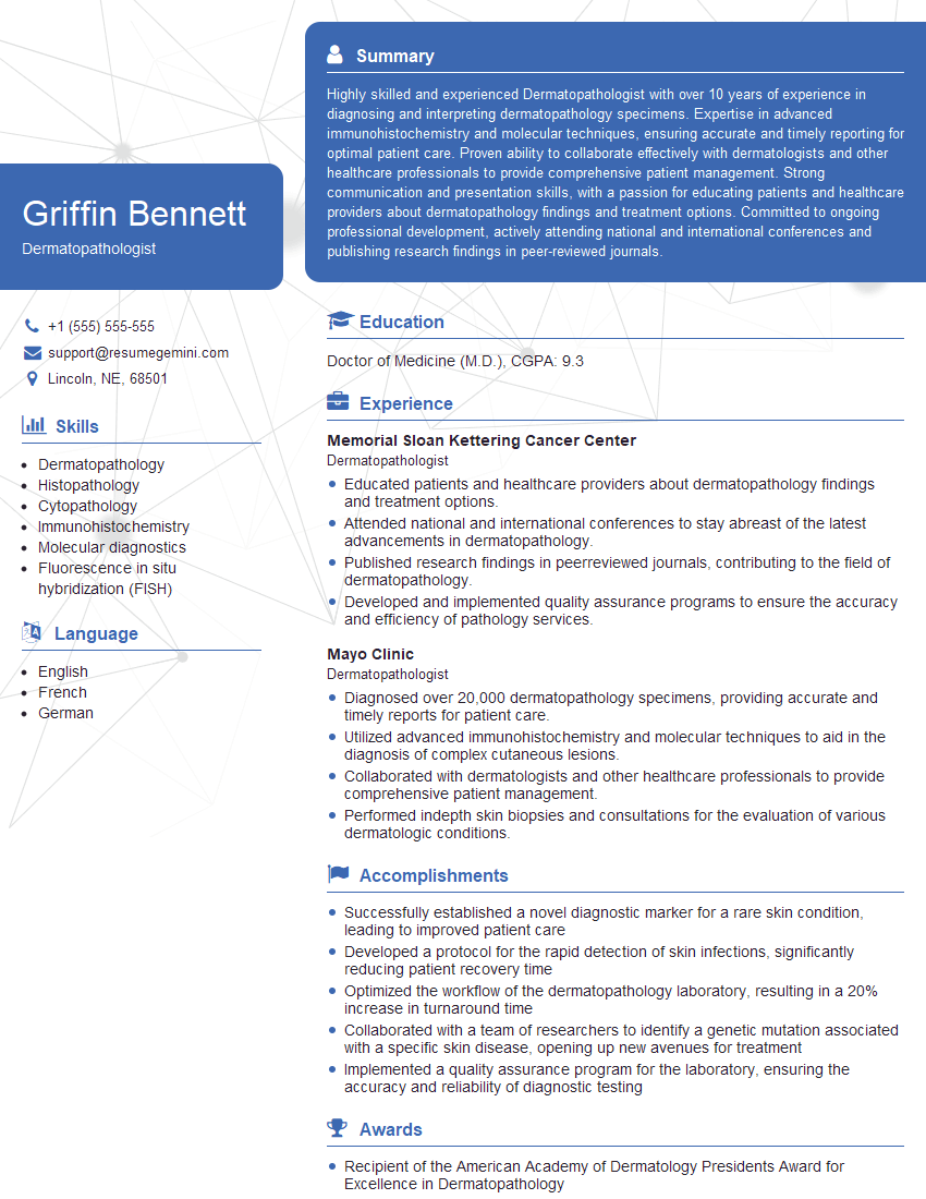
Ever felt underprepared for that crucial job interview? Or perhaps you’ve landed the interview but struggled to articulate your skills and experiences effectively? Fear not! We’ve got you covered. In this blog post, we’re diving deep into the Dermatopathologist interview questions that you’re most likely to encounter. But that’s not all. We’ll also provide expert insights into the key responsibilities of a Dermatopathologist so you can tailor your answers to impress potential employers.
Acing the interview is crucial, but landing one requires a compelling resume that gets you noticed. Crafting a professional document that highlights your skills and experience is the first step toward interview success. ResumeGemini can help you build a standout resume that gets you called in for that dream job.
Essential Interview Questions For Dermatopathologist
1. What are the key features that distinguish a melanocytic nevus from a melanoma?
- Melanocytic nevi are symmetric, with a well-demarcated border and uniform pigmentation.
- Melanomas are often asymmetric, with an irregular border and variegated pigmentation.
- Melanocytic nevi usually show a nested or compound growth pattern, while melanomas may exhibit a pagetoid or lentiginous growth pattern.
- Melanoma cells often show nuclear atypia, mitotic figures, and ulceration.
2. Describe the histologic features of a basal cell carcinoma.
Basaloid pattern
- Cells are arranged in small nests or cords, resembling the basal layer of the epidermis.
- Nests may be surrounded by a palisading layer of peripheral cells.
Peripheral palisading
- Cells at the periphery of nests are taller and more columnar, with their nuclei oriented perpendicular to the basement membrane.
- This feature is most prominent in early, well-differentiated lesions.
3. How do you differentiate between a seborrheic keratosis and an actinic keratosis?
- Seborrheic keratoses are usually larger and more elevated than actinic keratoses.
- Seborrheic keratoses have a characteristic “stuck-on” appearance, while actinic keratoses are often more scaly and crusty.
- On histologic examination, seborrheic keratoses show a thickening of the epidermis with a warty appearance, while actinic keratoses show atypical keratinocytes and solar elastosis.
4. Discuss the different types of cutaneous lymphomas (CL).
- Mycosis fungoides: The most common type of CL, presenting as scaly, erythematous patches or plaques that may evolve into tumors.
- Sézary syndrome: A leukemic variant of mycosis fungoides, characterized by erythroderma and circulating atypical lymphocytes in the peripheral blood.
- Follicular lymphoma: May present as solitary or multiple, firm, painless nodules in the skin, lymph nodes, or other organs.
- Peripheral T-cell lymphoma, not otherwise specified: A heterogeneous group of aggressive lymphomas that can involve the skin and other organs.
- Cutaneous B-cell lymphoma: A rare type of CL, presenting as solitary or multiple, painless nodules or tumors in the skin.
5. How would you approach the diagnosis of a suspected autoimmune blistering disorder?
- Obtain a detailed history, including a description of the lesions and any associated symptoms.
- Perform a physical examination, noting the distribution and morphology of the lesions.
- Order laboratory tests, including a blood count, chemistry panel, and autoimmune antibody profile.
- Consider performing a skin biopsy for histologic and immunofluorescence studies.
6. What are the key histologic features of a discoid lupus erythematosus (DLE) lesion?
- Interface dermatitis with vacuolar degeneration of the basal layer.
- Thickening of the basement membrane zone.
- Dermal fibrosis with a lichenoid infiltrate.
7. Describe the histologic findings in a bullous pemphigoid (BP) lesion.
Subepidermal blistering
- Blisters form beneath the epidermis, separating the epidermis from the dermis.
- The roof of the blister is composed of epidermis, while the base is composed of dermis.
Eosinophilic infiltrate
- Large numbers of eosinophils are present in the blister fluid and the underlying dermis.
- Eosinophils release cytotoxic granules that damage the basement membrane zone.
8. What are the most common cutaneous manifestations of syphilis?
- Primary syphilis: Characterized by a painless, indurated ulcer (chancre) at the site of inoculation.
- Secondary syphilis: Occurs weeks to months after the chancre heals and may include a maculopapular rash, lymphadenopathy, and fever.
- Tertiary syphilis: Develops years after the initial infection and may involve the skin, bones, cardiovascular system, and central nervous system.
9. How would you diagnose a case of pityriasis rosea?
- Obtain a detailed history, including the time course of the eruption and any associated symptoms.
- Perform a physical examination, noting the distribution and morphology of the lesions.
- Consider performing a skin biopsy if the diagnosis is uncertain.
10. What are the key histologic features of a lichen planus lesion?
- Hyperkeratosis
- Acanthosis
- Liquefactive degeneration of the basal layer
- Band-like infiltrate of lymphocytes in the upper dermis
- Pigmentation incontinence
Interviewers often ask about specific skills and experiences. With ResumeGemini‘s customizable templates, you can tailor your resume to showcase the skills most relevant to the position, making a powerful first impression. Also check out Resume Template specially tailored for Dermatopathologist.
Career Expert Tips:
- Ace those interviews! Prepare effectively by reviewing the Top 50 Most Common Interview Questions on ResumeGemini.
- Navigate your job search with confidence! Explore a wide range of Career Tips on ResumeGemini. Learn about common challenges and recommendations to overcome them.
- Craft the perfect resume! Master the Art of Resume Writing with ResumeGemini’s guide. Showcase your unique qualifications and achievements effectively.
- Great Savings With New Year Deals and Discounts! In 2025, boost your job search and build your dream resume with ResumeGemini’s ATS optimized templates.
Researching the company and tailoring your answers is essential. Once you have a clear understanding of the Dermatopathologist‘s requirements, you can use ResumeGemini to adjust your resume to perfectly match the job description.
Key Job Responsibilities
Dermatopathologists are highly specialized physicians responsible for examining cutaneous and peripheral nerve biopsies to diagnose skin disorders. Key responsibilities include:1. Microscopic Examination and Diagnosis
Examine skin biopsies under a microscope to identify abnormal cellular and tissue structures.
- Determine the type and severity of skin lesions.
- Provide histopathological interpretations to assist in diagnosis and patient management.
2. Consultation and Collaboration
Consult with dermatologists, medical oncologists, and other specialists to provide expert opinions on skin conditions.
- Participate in patient consultations to discuss findings and recommend treatment plans.
- Collaborate on research projects to advance the understanding and management of skin disorders.
3. Research and Education
Conduct or participate in research studies to enhance diagnostic techniques and treatment strategies.
- Publish findings in peer-reviewed journals and present them at conferences.
- Educate medical students, residents, and other healthcare professionals on dermatopathology.
4. Quality Assurance and Improvement
Maintain high standards of accuracy in diagnostic interpretations.
- Implement quality control measures to ensure reliable and timely reporting of results.
- Participate in proficiency testing programs to evaluate diagnostic skills.
Interview Tips
Preparing for a dermatopathology interview requires thorough research and practice. Here are some key tips:Study the job description carefully and identify the essential qualifications. Understand the specific responsibilities and expectations of the role.
1. Research the Organization and Industry
Research the hospital, clinic, or institution you are applying to. Learn about their mission, values, and areas of expertise.
- Demonstrate your knowledge of the organization’s history, research, and clinical programs.
- Show that your values and career goals align with the organization’s objectives.
2. Practice Your Technical Skills
Review the key techniques and procedures used in dermatopathology, such as biopsy interpretation, immunohistochemistry staining, and molecular diagnostics.
- Be prepared to discuss specific cases where you have demonstrated your technical expertise.
- Consider bringing a portfolio of your work to showcase your diagnostic skills.
3. Emphasize Your Communication and Collaboration Skills
Dermatopathologists interact with a diverse range of healthcare professionals. Highlight your ability to communicate effectively, build relationships, and collaborate on complex cases.
- Share examples where you have successfully consulted with clinicians and patients.
- Describe your experience in presenting complex medical information in a clear and understandable manner.
4. Demonstrate Your Passion for Dermatopathology
Convey your enthusiasm for the field and explain why you are passionate about dermatopathology.
- Discuss your research interests, publications, or volunteer experiences in the field.
- Show that you are actively engaged in professional development opportunities and stay updated on the latest advancements in dermatopathology.
Next Step:
Armed with this knowledge, you’re now well-equipped to tackle the Dermatopathologist interview with confidence. Remember, a well-crafted resume is your first impression. Take the time to tailor your resume to highlight your relevant skills and experiences. And don’t forget to practice your answers to common interview questions. With a little preparation, you’ll be on your way to landing your dream job. So what are you waiting for? Start building your resume and start applying! Build an amazing resume with ResumeGemini.
