Are you gearing up for an interview for a Histopathologist position? Whether you’re a seasoned professional or just stepping into the role, understanding what’s expected can make all the difference. In this blog, we dive deep into the essential interview questions for Histopathologist and break down the key responsibilities of the role. By exploring these insights, you’ll gain a clearer picture of what employers are looking for and how you can stand out. Read on to equip yourself with the knowledge and confidence needed to ace your next interview and land your dream job!
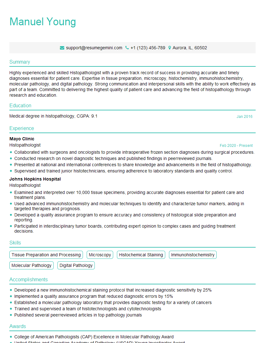
Acing the interview is crucial, but landing one requires a compelling resume that gets you noticed. Crafting a professional document that highlights your skills and experience is the first step toward interview success. ResumeGemini can help you build a standout resume that gets you called in for that dream job.
Essential Interview Questions For Histopathologist
1. How would you approach the diagnosis of a small round blue cell tumor?
- Histologic features: evaluate size, shape, and chromatin pattern of the cells, as well as the presence of rosettes, Homer-Wright rosettes, or other characteristic features.
- Immunohistochemistry: use a panel of antibodies to differentiate between different types of small round blue cell tumors, such as lymphoma, neuroblastoma, and Ewing sarcoma.
- Molecular studies: perform molecular testing, such as FISH or PCR, to identify specific genetic alterations that can help in the diagnosis.
- Clinical history and imaging studies: consider the patient’s age, symptoms, and imaging findings to narrow down the differential diagnosis.
2. What are the key microscopic features that distinguish between squamous cell carcinoma and basal cell carcinoma?
Microscopic features of squamous cell carcinoma:
- Intercellular bridges
- Keratin pearls
- Dyskeratotic cells
Microscopic features of basal cell carcinoma:
- Peripheral palisading of basaloid cells
- Retraction artifacts
- Cystic change
3. How would you assess the adequacy of a biopsy specimen for the diagnosis of an infectious disease?
- Evaluate the size and depth of the biopsy specimen.
- Assess the cellularity and presence of inflammatory cells.
- Identify the presence of the suspected infectious agent using appropriate stains or molecular techniques.
- Consider the clinical history and imaging findings to determine if the biopsy is likely to be representative of the underlying pathology.
4. What are the different types of melanocytic nevi, and how do you differentiate between them?
- Junctional nevus: Nevus cells are located at the dermal-epidermal junction.
- Compound nevus: Nevus cells are present in both the dermis and epidermis.
- Intradermal nevus: Nevus cells are located solely in the dermis.
- Dysplastic nevus: Atypical melanocytic nevus that has some features of melanoma, such as architectural disarray and pleomorphic nuclei. Diffuse atypia with melanocytes extending beyond the rete ridges.
- Blue nevus: Nevus cells contain melanin with a bluish hue.
5. How would you approach the diagnosis of a soft tissue sarcoma?
- Histologic features: assess the cellularity, architecture, and presence of specific histologic patterns.
- Immunohistochemistry: use a panel of antibodies to differentiate between different types of soft tissue sarcomas, such as leiomyosarcoma, liposarcoma, and synovial sarcoma.
- Molecular studies: perform molecular testing, such as FISH or PCR, to identify specific genetic alterations that can help in the diagnosis.
- Clinical history and imaging studies: consider the patient’s age, symptoms, and imaging findings to narrow down the differential diagnosis.
6. What are the different types of grading systems used for breast cancer, and how do they compare?
- Bloom-Richardson grading system: Based on tubule formation, nuclear grade, and mitotic count.
- Nottingham grading system: Similar to the Bloom-Richardson system, but also includes tumor size and lymphatic invasion.
- Scarff-Bloom-Richardson grading system: Focuses on nuclear grade and mitotic count.
- Elston-Ellis grading system: Assesses tumor size, nodal status, and histologic grade.
7. How would you assess the severity of chronic hepatitis?
- Histologic features: Grade the degree of inflammation, fibrosis, and necrosis.
- Immunohistochemistry: Assess the expression of specific markers, such as hepatitis B surface antigen or hepatitis C virus RNA, to determine the etiology of the hepatitis.
- Clinical history and laboratory tests: Consider the patient’s symptoms, liver function tests, and viral load to assess the severity of the disease.
8. What are the different types of amyloid, and how do you differentiate between them?
- AL amyloid: Derived from immunoglobulin light chains.
- AA amyloid: Derived from serum amyloid A protein.
- ATTR amyloid: Derived from transthyretin.
- Aβ amyloid: Derived from amyloid beta protein.
- PrP amyloid: Derived from prion protein.
9. How would you approach the diagnosis of a renal biopsy?
- Light microscopy: Assess the glomerular, tubular, and interstitial compartments for abnormalities.
- Immunofluorescence: Use antibodies to identify specific proteins, such as immunoglobulins or complement components, to help determine the etiology of the renal disease.
- Electron microscopy: Provides ultrastructural details that can help in the diagnosis of certain renal diseases, such as minimal change disease or membranous nephropathy.
- Clinical history and laboratory tests: Consider the patient’s symptoms, urine analysis, and blood tests to narrow down the differential diagnosis.
10. What are the different types of immunofluorescence patterns seen in glomerulonephritis, and what do they indicate?
- Granular pattern: Deposition of immune complexes in the glomerulus, seen in diseases such as lupus nephritis and post-infectious glomerulonephritis.
- Linear pattern: Deposition of antibodies along the glomerular basement membrane, seen in diseases such as Goodpasture’s syndrome and anti-GBM disease.
- Mesangial pattern: Deposition of immune complexes in the mesangium, seen in diseases such as IgA nephropathy and diabetic nephropathy.
- Membranous pattern: Deposition of antibodies along the glomerular capillary walls, seen in diseases such as membranous nephropathy.
Interviewers often ask about specific skills and experiences. With ResumeGemini‘s customizable templates, you can tailor your resume to showcase the skills most relevant to the position, making a powerful first impression. Also check out Resume Template specially tailored for Histopathologist.
Career Expert Tips:
- Ace those interviews! Prepare effectively by reviewing the Top 50 Most Common Interview Questions on ResumeGemini.
- Navigate your job search with confidence! Explore a wide range of Career Tips on ResumeGemini. Learn about common challenges and recommendations to overcome them.
- Craft the perfect resume! Master the Art of Resume Writing with ResumeGemini’s guide. Showcase your unique qualifications and achievements effectively.
- Great Savings With New Year Deals and Discounts! In 2025, boost your job search and build your dream resume with ResumeGemini’s ATS optimized templates.
Researching the company and tailoring your answers is essential. Once you have a clear understanding of the Histopathologist‘s requirements, you can use ResumeGemini to adjust your resume to perfectly match the job description.
Key Job Responsibilities
Histopathologists are medical professionals who diagnose and study diseases of the tissues. They play a critical role in healthcare by providing essential information to clinicians for patient care and treatment planning.
1. Tissue Examination and Analysis
Examining and analyzing tissue samples under a microscope to identify abnormal cells and structures.
- Performing biopsies and preparing tissue samples for examination.
- Using various staining techniques to highlight specific tissue components.
2. Diagnosis and Reporting
Diagnosing diseases based on the microscopic examination of tissue samples and providing detailed reports to clinicians.
- Identifying and classifying different types of diseases, such as cancer, infections, and autoimmune disorders.
- Documenting findings in medical records and issuing reports that include diagnoses, prognoses, and recommendations for treatment.
3. Consultation and Collaboration
Consulting with clinicians, surgeons, and other healthcare professionals to discuss patient cases and provide expert opinions.
- Providing guidance on the interpretation of test results and recommending follow-up procedures.
- Participating in multidisciplinary teams to develop and implement treatment plans.
4. Research and Development
Conducting research to improve diagnostic techniques and advance the field of histopathology.
- Developing new staining methods and molecular assays.
- Investigating the relationship between tissue changes and disease progression.
Interview Tips
Preparing for a histopathologist interview requires a combination of technical knowledge, communication skills, and professional demeanor. Here are some tips to help you ace your interview:
1. Research the Position and Organization
Familiarize yourself with the specific job requirements and the organization’s mission and values.
- Review the job description thoroughly and identify the key responsibilities and qualifications.
- Visit the organization’s website and LinkedIn page to learn about their culture and recent developments.
2. Highlight Your Technical Skills
Emphasize your expertise in histopathology techniques, including tissue preparation, microscopy, and diagnostic interpretation.
- Provide specific examples of complex cases you have diagnosed and the impact of your findings on patient care.
- Discuss your experience with different staining methods, molecular assays, and research projects.
3. Demonstrate Strong Communication Skills
Showcasing your ability to communicate effectively is crucial for histopathologists who often collaborate with other healthcare professionals and patients.
- Prepare clear and concise presentations that highlight your diagnostic findings and recommendations.
- Practice discussing complex medical concepts in a way that is understandable to both clinicians and patients.
4. Convey Professionalism and Enthusiasm
Dress professionally, arrive on time for the interview, and maintain a positive and enthusiastic attitude throughout the process.
- Show respect for the interviewers and the organization by asking thoughtful questions and listening attentively.
- Express your passion for histopathology and your commitment to providing high-quality patient care.
Next Step:
Now that you’re armed with the knowledge of Histopathologist interview questions and responsibilities, it’s time to take the next step. Build or refine your resume to highlight your skills and experiences that align with this role. Don’t be afraid to tailor your resume to each specific job application. Finally, start applying for Histopathologist positions with confidence. Remember, preparation is key, and with the right approach, you’ll be well on your way to landing your dream job. Build an amazing resume with ResumeGemini
