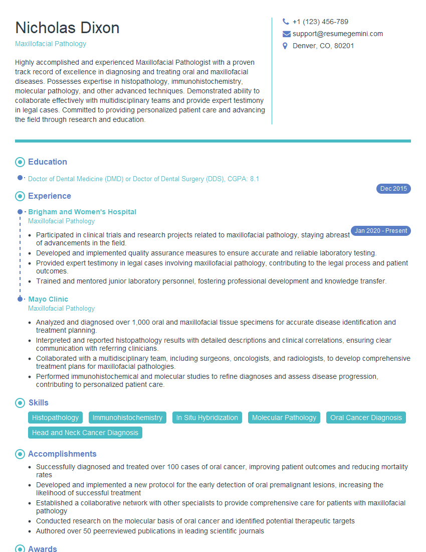
Are you gearing up for a career in Maxillofacial Pathology? Feeling nervous about the interview questions that might come your way? Don’t worry, you’re in the right place. In this blog post, we’ll dive deep into the most common interview questions for Maxillofacial Pathology and provide you with expert-backed answers. We’ll also explore the key responsibilities of this role so you can tailor your responses to showcase your perfect fit.
Acing the interview is crucial, but landing one requires a compelling resume that gets you noticed. Crafting a professional document that highlights your skills and experience is the first step toward interview success. ResumeGemini can help you build a standout resume that gets you called in for that dream job.
Essential Interview Questions For Maxillofacial Pathology
1. What is the difference between odontogenic cysts and non-odontogenic cysts in the jaws?
answer:
- Odontogenic cysts arise from the remnants of the dental lamina, while non-odontogenic cysts arise from non-dental tissues.
- Odontogenic cysts are usually lined by epithelium, while non-odontogenic cysts are lined by various types of tissue, such as respiratory epithelium, salivary gland tissue, or mucous membrane.
- Odontogenic cysts are often associated with impacted teeth, while non-odontogenic cysts are not.
2. What are the key microscopic features that distinguish between squamous cell carcinoma and verrucous carcinoma of the oral cavity?
answer:
Microscopic features of squamous cell carcinoma:
- Invasive growth pattern
- Atypical cells with large, hyperchromatic nuclei
- Keratin pearl formation
- Intercellular bridges
Microscopic features of verrucous carcinoma:
- Exophytic growth pattern
- Well-differentiated cells with small, round nuclei
- No keratin pearl formation
- Wide intercellular bridges
3. How do you differentiate between benign and malignant salivary gland tumors?
answer:
- Clinical features: Benign tumors are usually slow-growing and painless, while malignant tumors may grow rapidly and cause pain.
- Imaging findings: Benign tumors typically have well-defined borders on imaging studies, while malignant tumors may have irregular borders.
- Microscopic features: Benign tumors have well-differentiated cells and a low mitotic index, while malignant tumors have poorly differentiated cells and a high mitotic index.
4. What are the histologic features of a Warthin’s tumor?
answer:
- Bilobed architecture
- Cystic and papillary areas
- Oncocytic (eosinophilic) epithelial cells
- Lymphoid stroma with germinal centers
5. What is the microscopic differential diagnosis of a spindle cell tumor of the oral cavity?
answer:
- Schwannoma
- Neurofibroma
- Leiomyoma
- Fibrosarcoma
- Malignant peripheral nerve sheath tumor
6. How do you assess the adequacy of a biopsy specimen for a suspected case of oral cancer?
answer:
- Ensure that the specimen includes representative areas of the lesion, including the invasive front.
- Check that the specimen is of sufficient size to allow for proper histologic evaluation.
- Orient the specimen correctly to facilitate histologic interpretation.
- Fix the specimen in an appropriate fixative to preserve tissue morphology.
7. What are the immunohistochemical markers that can be used to differentiate between osteosarcoma and Ewing’s sarcoma?
answer:
- Osteosarcoma: S-100 protein, vimentin, alkaline phosphatase
- Ewing’s sarcoma: CD99, MIC2, FLI-1
8. How do you interpret a positive immunohistochemical stain for p16 in a biopsy specimen of oral squamous cell carcinoma?
answer:
- A positive p16 stain indicates that the tumor cells have lost function of the RB1 gene.
- Loss of RB1 function is associated with increased cell proliferation and tumor progression.
- A positive p16 stain is a prognostic indicator of poor survival in oral squamous cell carcinoma.
9. What are the different types of salivary gland neoplasms?
answer:
- Benign: Pleomorphic adenoma, Warthin’s tumor, basal cell adenoma, canalicular adenoma, mucoepidermoid adenoma
- Malignant: Mucoepidermoid carcinoma, adenoid cystic carcinoma, acinic cell carcinoma, salivary duct carcinoma, undifferentiated carcinoma
10. What is the role of a maxillofacial pathologist in the management of patients with head and neck cancer?
answer:
- Provide a definitive diagnosis of head and neck cancers.
- Determine the stage and grade of head and neck cancers.
- Monitor patients with head and neck cancer for recurrence or progression.
- Participate in clinical trials and research on head and neck cancer.
- Educate patients and their families about head and neck cancer.
Interviewers often ask about specific skills and experiences. With ResumeGemini‘s customizable templates, you can tailor your resume to showcase the skills most relevant to the position, making a powerful first impression. Also check out Resume Template specially tailored for Maxillofacial Pathology.
Career Expert Tips:
- Ace those interviews! Prepare effectively by reviewing the Top 50 Most Common Interview Questions on ResumeGemini.
- Navigate your job search with confidence! Explore a wide range of Career Tips on ResumeGemini. Learn about common challenges and recommendations to overcome them.
- Craft the perfect resume! Master the Art of Resume Writing with ResumeGemini’s guide. Showcase your unique qualifications and achievements effectively.
- Great Savings With New Year Deals and Discounts! In 2025, boost your job search and build your dream resume with ResumeGemini’s ATS optimized templates.
Researching the company and tailoring your answers is essential. Once you have a clear understanding of the Maxillofacial Pathology‘s requirements, you can use ResumeGemini to adjust your resume to perfectly match the job description.
Key Job Responsibilities
Maxillofacial Pathologists are highly specialized medical professionals who play a crucial role in diagnosing and managing diseases of the mouth, jaws, and associated tissues. Their responsibilities encompass a wide range of tasks, including:
1. Diagnostic Procedures
Examining tissue samples under a microscope to identify abnormalities and determine the nature of the disease. This involves preparing and staining the tissue, using advanced techniques such as immunohistochemistry and molecular diagnostics.
2. Consultation and Collaboration
Providing expert opinions to other healthcare professionals, including dentists, oral surgeons, and oncologists, to assist in the diagnosis, treatment planning, and management of patients with oral and maxillofacial diseases.
3. Research and Education
Conducting original research to advance the understanding of oral and maxillofacial diseases. This may involve studying the molecular mechanisms of disease, developing new diagnostic methods, or evaluating the efficacy of treatments.
4. Management and Administration
Supervising laboratory staff, ensuring quality control, and managing the overall operation of the pathology laboratory. This includes maintaining equipment, ordering supplies, and ensuring compliance with regulatory standards.
Interview Tips
Preparing for an interview for a Maxillofacial Pathology position requires thorough research and careful consideration of the specific requirements of the role. Here are some key tips to help you ace the interview:
1. Research the Organization and Position
Familiarize yourself with the organization’s mission, values, and current research focus. Research the specific job responsibilities and qualifications required for the position.
2. Highlight Your Skills and Experience
Tailor your resume and cover letter to emphasize the skills and experience that are most relevant to the job description. Quantify your accomplishments and provide specific examples of your contributions.
3. Practice Your Answers
Anticipate common interview questions and prepare thoughtful answers that demonstrate your knowledge, skills, and enthusiasm for the field. Practice answering questions out loud to improve your delivery.
4. Be Prepared to Discuss Your Research
If you have conducted research in the field of Maxillofacial Pathology, be prepared to discuss your findings and how they contribute to the understanding or management of oral and maxillofacial diseases.
5. Dress Professionally and Arrive on Time
First impressions matter. Dress appropriately for a professional setting and arrive on time for your interview. Punctuality and a polished appearance demonstrate respect for the interviewers and the organization.
Next Step:
Armed with this knowledge, you’re now well-equipped to tackle the Maxillofacial Pathology interview with confidence. Remember, a well-crafted resume is your first impression. Take the time to tailor your resume to highlight your relevant skills and experiences. And don’t forget to practice your answers to common interview questions. With a little preparation, you’ll be on your way to landing your dream job. So what are you waiting for? Start building your resume and start applying! Build an amazing resume with ResumeGemini.
