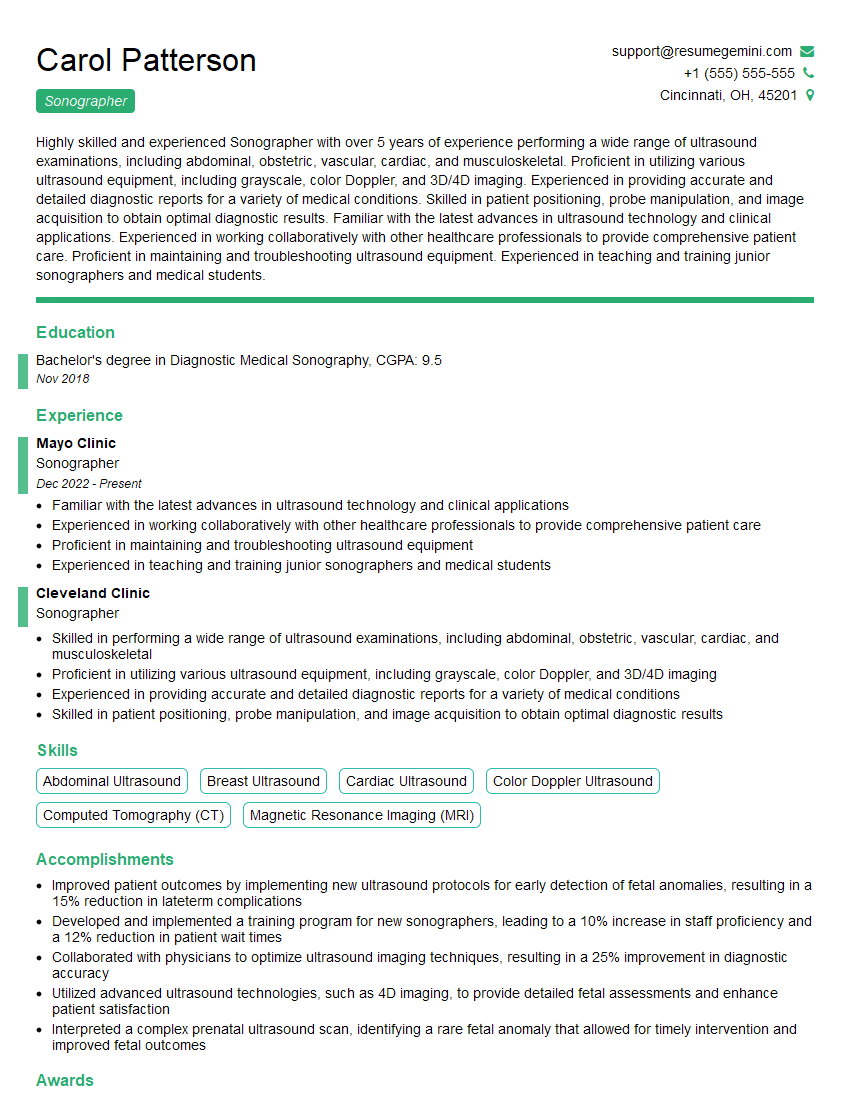
Are you gearing up for a career shift or aiming to ace your next interview? Look no further! We’ve curated a comprehensive guide to help you crack the interview for the coveted Sonographer position. From understanding the key responsibilities to mastering the most commonly asked questions, this blog has you covered. So, buckle up and let’s embark on this journey together
Acing the interview is crucial, but landing one requires a compelling resume that gets you noticed. Crafting a professional document that highlights your skills and experience is the first step toward interview success. ResumeGemini can help you build a standout resume that gets you called in for that dream job.
Essential Interview Questions For Sonographer
1. What are the key physiological principles underlying ultrasound imaging?
In ultrasound imaging, high-frequency sound waves are emitted into the body. These waves interact with tissues and organs, and the resulting echoes are used to create images. The key physiological principles underlying ultrasound imaging include:
- Acoustic impedance: The acoustic impedance of a tissue is a measure of its resistance to the passage of sound waves. Different tissues have different acoustic impedances, which is why they reflect sound waves differently.
- Attenuation: Sound waves are attenuated, or weakened, as they pass through tissues. The attenuation of sound waves is dependent on the frequency of the waves, the type of tissue, and the distance the waves travel.
- Scattering: Sound waves are scattered by particles in tissues. The scattering of sound waves is dependent on the size, shape, and density of the particles.
2. Describe the different types of ultrasound transducers and their applications.
There are two main types of ultrasound transducers: linear and curved.
Linear transducers
- Produce rectangular images
- Best for superficial imaging, such as the abdomen, breast, and thyroid
Curved transducers
- Produce sector-shaped images
- Best for deep imaging, such as the heart, liver, and kidneys
3. Explain the concept of Doppler ultrasound and its clinical applications.
Doppler ultrasound is a technique that uses the Doppler effect to measure the velocity of blood flow. The Doppler effect is a change in the frequency of a wave that occurs when the source of the wave is moving relative to the observer. In Doppler ultrasound, the source of the wave is the ultrasound transducer and the observer is the patient. When blood is flowing, it causes the ultrasound waves to change frequency. The amount of frequency change is proportional to the velocity of the blood flow.
Doppler ultrasound has a variety of clinical applications, including:
- Evaluating blood flow in the heart and blood vessels
- Detecting blood clots
- Monitoring the development of fetuses
4. What are the most common artifacts in ultrasound imaging and how can they be minimized?
The most common artifacts in ultrasound imaging include:
- Acoustic shadowing: This occurs when a dense tissue, such as bone, blocks the transmission of sound waves. The area behind the dense tissue will appear dark on the ultrasound image.
- Reverberation: This occurs when sound waves bounce back and forth between two dense tissues, such as bone and muscle. The reverberation will appear as a series of parallel lines on the ultrasound image.
- Side lobes: These are artifacts that occur when sound waves are diffracted around a small object. Side lobes can appear as bright or dark lines on the ultrasound image.
Artifacts can be minimized by using the appropriate transducer, adjusting the gain and frequency settings, and using techniques such as harmonic imaging and compound imaging.
5. Describe the role of ultrasound in the diagnosis and management of cardiovascular disease.
Ultrasound is a valuable tool for the diagnosis and management of cardiovascular disease. It can be used to:
- Evaluate the structure and function of the heart
- Detect heart defects
- Monitor the progression of cardiovascular disease
- Guide interventional procedures
Ultrasound is a non-invasive and relatively inexpensive imaging modality, making it ideal for screening and monitoring patients with cardiovascular disease.
6. What are the advantages and disadvantages of ultrasound imaging compared to other imaging modalities such as CT and MRI?
Advantages of ultrasound imaging:
- Non-invasive: Ultrasound does not involve the use of ionizing radiation, making it a safe imaging modality for pregnant women and children.
- Real-time imaging: Ultrasound allows for real-time imaging, which is useful for evaluating moving structures such as the heart and blood vessels.
- Cost-effective: Ultrasound is a relatively inexpensive imaging modality, making it accessible to a wider range of patients.
Disadvantages of ultrasound imaging:
- Limited depth of penetration: Ultrasound waves cannot penetrate deeply into the body, making it difficult to image deep structures such as the brain and lungs.
- Operator-dependent: The quality of ultrasound images is highly dependent on the skill and experience of the sonographer.
- Artifacts: Ultrasound images can be affected by artifacts, which can make it difficult to interpret the images.
7. What are the ethical considerations in ultrasound imaging?
Ultrasound is a powerful imaging tool, but it is important to use it ethically. Some of the ethical considerations in ultrasound imaging include:
- Patient consent: Patients should be fully informed about the risks and benefits of ultrasound imaging before they consent to the procedure.
- Confidentiality: Ultrasound images and information should be kept confidential and only shared with those who need to know.
- Respect for the patient’s body: Sonographers should always respect the patient’s body and privacy.
8. What are the future trends in ultrasound imaging?
The future of ultrasound imaging is bright. Some of the trends that are likely to shape the future of ultrasound imaging include:
- Three-dimensional ultrasound: Three-dimensional ultrasound allows for the creation of three-dimensional images of organs and tissues.
- Contrast-enhanced ultrasound: Contrast-enhanced ultrasound uses contrast agents to improve the visualization of blood flow and other structures.
- Molecular imaging: Molecular imaging uses ultrasound to detect and image molecular markers of disease.
9. How do you stay up-to-date on the latest advances in ultrasound imaging?
I stay up-to-date on the latest advances in ultrasound imaging by reading journals, attending conferences, and participating in continuing education courses.
- Journals: I read a variety of ultrasound journals, including the Journal of Ultrasound in Medicine, Ultrasound Quarterly, and the International Journal of Ultrasonography.
- Conferences: I attend national and international ultrasound conferences, such as the annual meeting of the American Institute of Ultrasound in Medicine.
- Continuing education courses: I participate in continuing education courses offered by my hospital and by professional organizations such as the American Registry for Diagnostic Medical Sonographers.
10. What are your greatest strengths and weaknesses as a sonographer?
Strengths:
- I have a strong understanding of the principles of ultrasound imaging.
- I am proficient in using a variety of ultrasound transducers and imaging techniques.
- I am able to obtain high-quality images that are essential for accurate diagnosis.
- I am comfortable working with patients from all walks of life.
Weaknesses:
- I am still relatively new to the field of ultrasound imaging.
- I am not yet proficient in all of the advanced ultrasound imaging techniques.
- I am sometimes uncomfortable working with patients who are anxious or uncooperative.
Interviewers often ask about specific skills and experiences. With ResumeGemini‘s customizable templates, you can tailor your resume to showcase the skills most relevant to the position, making a powerful first impression. Also check out Resume Template specially tailored for Sonographer.
Career Expert Tips:
- Ace those interviews! Prepare effectively by reviewing the Top 50 Most Common Interview Questions on ResumeGemini.
- Navigate your job search with confidence! Explore a wide range of Career Tips on ResumeGemini. Learn about common challenges and recommendations to overcome them.
- Craft the perfect resume! Master the Art of Resume Writing with ResumeGemini’s guide. Showcase your unique qualifications and achievements effectively.
- Great Savings With New Year Deals and Discounts! In 2025, boost your job search and build your dream resume with ResumeGemini’s ATS optimized templates.
Researching the company and tailoring your answers is essential. Once you have a clear understanding of the Sonographer‘s requirements, you can use ResumeGemini to adjust your resume to perfectly match the job description.
Key Job Responsibilities
Sonographers play a critical role in medical imaging by using ultrasound technology to capture images of internal organs and structures. Their key job responsibilities encompass a range of technical and patient-centered tasks.
1. Patient Care and Preparation
Interact with patients, explain procedures, and obtain informed consent.
- Prepare patients for exams by providing clear instructions and positioning them appropriately.
- Maintain a respectful and professional demeanor throughout patient interactions.
2. Ultrasound Imaging Acquisition
Operate ultrasound equipment and acquire high-quality diagnostic images.
- Select appropriate imaging techniques and adjust imaging parameters to optimize image quality.
- Document and interpret ultrasound findings, identifying any abnormalities or areas of concern.
3. Image Analysis and Report Generation
Analyze images, determine appropriate measurements, and compose detailed reports.
- Use standardized reporting templates and accurately record all relevant findings.
- Collaborate with radiologists and other healthcare professionals to ensure accurate and timely image interpretation.
4. Equipment Maintenance and Quality Assurance
Ensure the proper functioning of ultrasound equipment and maintain high standards of image quality.
- Perform routine equipment checks, calibrations, and troubleshooting to maintain optimal performance.
- Implement quality assurance protocols to ensure the accuracy and consistency of ultrasound examinations.
5. Continuing Education and Professional Development
Stay abreast of advancements in ultrasound technology and best practices.
- Attend workshops, conferences, and training programs to enhance technical skills and knowledge.
- Maintain professional certifications and licenses to demonstrate ongoing professional competence.
Interview Tips
Preparing for a successful Sonographer interview involves researching the role, practicing your answers, and demonstrating your knowledge and skills.
1. Research the Role and Company
Thoroughly review the job description and company website to gain a comprehensive understanding of the position and the organization’s culture.
- Identify the key responsibilities and qualifications required for the role.
- Research the company’s mission, values, and recent news to demonstrate your interest and alignment with the organization.
2. Practice Your Answers
Prepare clear and concise answers to common interview questions related to your technical skills, patient care experience, and professional development.
- Use the STAR method (Situation, Task, Action, Result) when answering behavioral questions to provide specific examples of your abilities.
- Practice your responses out loud to improve your delivery and confidence.
3. Showcase Your Skills
Highlight your technical proficiency in ultrasound imaging, image interpretation, and report writing.
- Provide examples of challenging cases where you successfully identified and documented abnormalities.
- Share your experience in using advanced imaging techniques to enhance diagnostic accuracy.
4. Emphasize Your Patient Care Abilities
Emphasize your compassion, empathy, and ability to build rapport with patients.
- Describe strategies you use to create a comfortable and reassuring environment for patients.
- Share examples of how you have effectively communicated complex medical information to patients and their families.
5. Demonstrate Your Professionalism and Commitment
Convey your dedication to professionalism and continuous learning.
- Highlight your involvement in professional organizations, attendance at conferences, and pursuit of certifications.
- Express your enthusiasm for the field of sonography and your commitment to providing high-quality patient care.
Next Step:
Armed with this knowledge, you’re now well-equipped to tackle the Sonographer interview with confidence. Remember, preparation is key. So, start crafting your resume, highlighting your relevant skills and experiences. Don’t be afraid to tailor your application to each specific job posting. With the right approach and a bit of practice, you’ll be well on your way to landing your dream job. Build your resume now from scratch or optimize your existing resume with ResumeGemini. Wish you luck in your career journey!
