Are you gearing up for a career shift or aiming to ace your next interview? Look no further! We’ve curated a comprehensive guide to help you crack the interview for the coveted X Ray Technician position. From understanding the key responsibilities to mastering the most commonly asked questions, this blog has you covered. So, buckle up and let’s embark on this journey together
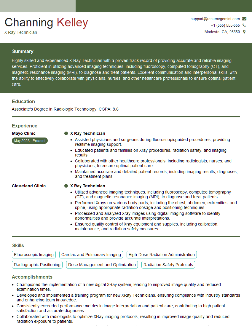
Acing the interview is crucial, but landing one requires a compelling resume that gets you noticed. Crafting a professional document that highlights your skills and experience is the first step toward interview success. ResumeGemini can help you build a standout resume that gets you called in for that dream job.
Essential Interview Questions For X Ray Technician
1. Describe the process of performing a chest X-ray.
The process of performing a chest X-ray involves several key steps:
- Patient preparation: Before the X-ray, the patient is asked to remove any metal objects or jewelry that could interfere with the image. The patient is positioned standing or sitting upright, with their chest facing the X-ray machine.
- Positioning: The X-ray technician positions the patient so that the area of interest is centered in the imaging field. This ensures that the desired anatomical structures are captured clearly.
- Exposure: The X-ray machine is then activated, emitting a short burst of radiation that passes through the patient’s chest. The radiation is absorbed by different tissues within the chest to varying degrees, creating an image on the X-ray detector.
- Image processing: The raw X-ray image is processed to improve its clarity and contrast. This may involve adjustments to brightness, contrast, and other image parameters to optimize the visualization of anatomical features.
2. What are the different types of X-ray equipment used in medical imaging?
X-ray tubes
- Stationary X-ray tubes: These tubes are permanently mounted within an X-ray machine and cannot be moved. They are typically used for general radiography and fluoroscopy.
- Mobile X-ray tubes: These tubes can be moved around on a wheeled stand, allowing for X-rays to be taken in various locations, such as at a patient’s bedside or in an operating room.
X-ray detectors
- Film-based detectors: Traditional X-rays use film as the detector to capture the radiation passing through the patient. The exposed film is then processed to produce a visible image.
- Digital detectors: Modern X-ray systems employ digital detectors, such as amorphous selenium (a-Se) or cesium iodide (CsI) panels. These detectors convert X-ray radiation into electronic signals, which are then processed and displayed as digital images.
3. How do you ensure the quality of X-ray images?
Maintaining the quality of X-ray images is crucial for accurate diagnosis. Key aspects of quality assurance include:
- Proper positioning of the patient and X-ray tube:
- Optimizing exposure parameters (kVp and mAs) to achieve the correct density and contrast.
- Regular calibration and maintenance of X-ray equipment to ensure accuracy and consistency.
- Quality control measures such as using phantoms and image evaluation tools to assess image quality and identify any potential issues.
- Following established protocols and guidelines for X-ray imaging to ensure consistency and adherence to best practices.
4. What are the radiation safety protocols you follow when working with X-ray equipment?
Radiation safety is paramount when working with X-ray equipment. I strictly adhere to the following protocols:
- Minimizing radiation exposure by using appropriate shielding, such as lead aprons and gloves, and maintaining a safe distance from the X-ray source.
- Following the principle of ALARA (As Low As Reasonably Achievable), ensuring that the lowest possible radiation dose is used to obtain the necessary diagnostic information.
- Regular monitoring of radiation exposure through the use of personal dosimeters to track cumulative radiation doses.
- Understanding and adhering to national and international radiation safety standards and guidelines.
5. Describe the role of contrast agents in X-ray imaging.
Contrast agents are substances used to enhance the visibility of specific anatomical structures or organs on X-ray images. In X-ray imaging, contrast agents can be administered orally, intravenously, or through other routes:
- Barium sulfate: This is commonly used to visualize the gastrointestinal tract, as it coats the lining of the organs and makes them more visible on X-rays.
- Iodine-based contrast agents: These are used for imaging blood vessels, the urinary tract, and other organs and tissues.
- Air or gas: Air can be introduced into body cavities, such as the abdomen or joints, to create contrast and improve visualization.
6. How do you handle patients who may be anxious or uncooperative during X-ray examinations?
Handling anxious or uncooperative patients during X-ray examinations requires a combination of empathy, communication skills, and technical expertise:
- Empathy and reassurance: It is important to understand and empathize with the patient’s fears or concerns. Reassurance and clear communication can help reduce anxiety.
- Effective communication: Clearly explaining the procedure, addressing any concerns, and providing step-by-step instructions can help the patient feel more comfortable and cooperative.
- Technical expertise: Proper positioning and immobilization techniques are crucial for obtaining clear and accurate X-ray images. Using appropriate restraints or sedation when necessary can help ensure patient cooperation and minimize motion artifacts.
7. What is your approach to optimizing patient positioning for different X-ray examinations?
Optimizing patient positioning is essential for obtaining high-quality X-ray images. My approach involves:
- Understanding the anatomical structures of interest and the desired view for each examination.
- Positioning the patient correctly to ensure that the area of interest is centered in the imaging field.
- Properly immobilizing the patient to minimize motion artifacts and improve image clarity.
- Using appropriate positioning aids, such as sandbags or foam blocks, to support the patient and maintain proper alignment.
8. How do you manage and reduce patient radiation dose during X-ray examinations?
Patient radiation dose management is a critical aspect of X-ray imaging. I employ several strategies to minimize dose while maintaining image quality:
- Using appropriate exposure parameters (kVp and mAs) based on the patient’s size and the anatomical area being imaged.
- Employing dose-reduction techniques, such as pulsed fluoroscopy and automatic exposure control (AEC).
- Utilizing shielding devices, such as lead aprons and thyroid collars, to protect sensitive areas from unnecessary radiation exposure.
- Regularly monitoring and evaluating patient radiation doses to ensure compliance with safety standards.
9. Describe the different imaging modalities used in X-ray technology and their applications.
X-ray technology encompasses various imaging modalities, each with unique applications:
- Plain film radiography: This is the most common type of X-ray imaging, used to visualize bones, lungs, and other anatomical structures.
- Fluoroscopy: This technique involves continuous X-ray imaging, allowing real-time visualization of moving structures, such as during gastrointestinal studies or fluoroscopic-guided procedures.
- Computed tomography (CT): CT scans combine multiple X-ray images to create cross-sectional images of the body, providing detailed views of internal structures.
- Digital subtraction angiography (DSA): DSA is a specialized X-ray technique used to visualize blood vessels by injecting a contrast agent and capturing images as the contrast flows through the vessels.
10. How do you stay up-to-date with advancements in X-ray technology and best practices?
To stay current with advancements in X-ray technology and best practices, I engage in ongoing professional development activities:
- Attending conferences, workshops, and webinars.
- Reading scientific journals and industry publications.
- Participating in continuing education programs and certifications.
- Seeking mentorship and guidance from experienced professionals.
- Collaborating with colleagues and sharing knowledge.
Interviewers often ask about specific skills and experiences. With ResumeGemini‘s customizable templates, you can tailor your resume to showcase the skills most relevant to the position, making a powerful first impression. Also check out Resume Template specially tailored for X Ray Technician.
Career Expert Tips:
- Ace those interviews! Prepare effectively by reviewing the Top 50 Most Common Interview Questions on ResumeGemini.
- Navigate your job search with confidence! Explore a wide range of Career Tips on ResumeGemini. Learn about common challenges and recommendations to overcome them.
- Craft the perfect resume! Master the Art of Resume Writing with ResumeGemini’s guide. Showcase your unique qualifications and achievements effectively.
- Great Savings With New Year Deals and Discounts! In 2025, boost your job search and build your dream resume with ResumeGemini’s ATS optimized templates.
Researching the company and tailoring your answers is essential. Once you have a clear understanding of the X Ray Technician‘s requirements, you can use ResumeGemini to adjust your resume to perfectly match the job description.
Key Job Responsibilities
X-ray technicians are responsible for taking medical x-ray images in hospitals, clinics, and other healthcare settings. They use specialized equipment to capture images of the human body, which are then used by physicians and other healthcare professionals to diagnose and treat medical conditions.
1. Patient care
X-ray technicians must be able to put patients at ease and make them comfortable during the x-ray procedure. They must also be able to communicate effectively with patients and answer their questions.
- Greet patients and explain the x-ray procedure.
- Position patients and ensure that they remain still during the x-ray.
- Answer patients’ questions and address their concerns.
- Provide comfort and support to patients during the x-ray procedure.
2. Image acquisition
X-ray technicians must be able to operate x-ray equipment safely and efficiently. They must also be able to adjust the equipment to produce high-quality images.
- Operate x-ray equipment and adjust settings to obtain optimal images.
- Position and expose patients to radiation to produce x-ray images.
- Process x-ray images and ensure that they are of good quality.
- Maintain and troubleshoot x-ray equipment.
3. Patient records
X-ray technicians must be able to maintain accurate patient records. They must also be able to track the progress of patients’ conditions.
- Maintain accurate patient records, including x-ray images.
- Track the progress of patients’ conditions by comparing x-ray images over time.
- Communicate with physicians and other healthcare professionals to discuss patients’ x-ray images.
4. Quality assurance
X-ray technicians must be able to participate in quality assurance programs to ensure that the x-ray department is providing high-quality care to patients.
- Participate in quality assurance programs to ensure the accuracy of x-ray images.
- Conduct regular inspections of x-ray equipment to ensure that it is functioning properly.
- Provide feedback to the quality assurance team and recommend improvements to the x-ray department.
Interview Tips
To prepare for your interview, you should research the job description and the company. You should also practice answering common interview questions. Here are some tips to help you ace your interview:
1. Dress professionally
First impressions matter, so it’s important to dress professionally for your interview. This means wearing a suit or other business attire. You should also make sure your hair is neat and your nails are clean.
2. Arrive on time
Punctuality is important, so make sure you arrive for your interview on time. If you’re running late, call the interviewer to let them know. It’s also a good idea to arrive a few minutes early so you can relax and collect your thoughts before the interview.
3. Be prepared to answer common interview questions
There are a few common interview questions that you should be prepared to answer. These questions include:
- Tell me about yourself.
- Why are you interested in this job?
- What are your strengths and weaknesses?
- What are your career goals?
- Why should we hire you?
You can prepare for these questions by practicing your answers ahead of time. You should also be prepared to give specific examples of your experience and skills.
4. Ask questions
At the end of the interview, the interviewer will likely ask if you have any questions. This is your opportunity to learn more about the job and the company. It’s also a good way to show the interviewer that you’re interested in the position.
Here are some good questions to ask:
- What are the biggest challenges facing the x-ray department?
- What are the opportunities for advancement within the company?
- What is the company culture like?
- What is the training program like for new employees?
5. Follow up
After the interview, it’s a good idea to follow up with the interviewer. This shows that you’re still interested in the position and that you appreciate their time. You can follow up by email or by phone.
In your follow-up, you should thank the interviewer for their time and reiterate your interest in the position. You can also use this opportunity to ask any additional questions that you may have.
Next Step:
Armed with this knowledge, you’re now well-equipped to tackle the X Ray Technician interview with confidence. Remember, a well-crafted resume is your first impression. Take the time to tailor your resume to highlight your relevant skills and experiences. And don’t forget to practice your answers to common interview questions. With a little preparation, you’ll be on your way to landing your dream job. So what are you waiting for? Start building your resume and start applying! Build an amazing resume with ResumeGemini.
